Introduction to Physical Examination
Introduction to Physical Examination. Dr. Gwen Hollaar Dr. Lanice Jones Dr. Robert Lee September 2006 Lao Project. Outline. General Observations Vital Signs General Approach to physical examination General surface anatomy Examination of Head and Neck Examination of Lymph nodes.
Share Presentation
Embed Code
Link
Download Presentation
- examination
- surface anatomy
- neck examination
- blood pressure
- neck examination thyroid
- surface anatomy anterior chest

sigrids + Follow
Download Presentation
Introduction to Physical Examination
An Image/Link below is provided (as is) to download presentation Download Policy: Content on the Website is provided to you AS IS for your information and personal use and may not be sold / licensed / shared on other websites without getting consent from its author. Content is provided to you AS IS for your information and personal use only. Download presentation by click this link. While downloading, if for some reason you are not able to download a presentation, the publisher may have deleted the file from their server. During download, if you can't get a presentation, the file might be deleted by the publisher.
Presentation Transcript
- Introduction to Physical Examination Dr. Gwen Hollaar Dr. Lanice Jones Dr. Robert Lee September 2006 Lao Project
- Outline • General Observations • Vital Signs • General Approach to physical examination • General surface anatomy • Examination of Head and Neck • Examination of Lymph nodes
- Physical Examination begins with observing the patient • Many observations can be made while the patient walks into the examination room or as you approach the patient • Level of Consciousness • Alert or decreased level of consciousness • Apparent State of Health: • Acutely ill or chronically ill (i.e. emaciated) • Signs of Distress • Sweating / Diaphoresis • Dyspnea / Wheeze / Cough • Pain • Posture, gait, or motor activity • Anxiety or Depression • Skin • Pale • Jaundice • Dress or hygiene
- Preparation / Draping Prior to beginning formal physical examination: • Explain to patient what you will be doing • Make sure patient feels comfortable and provide privacy • Ask patient to remove clothing so that you can do proper physical examination • Give patient sheet/gown to cover herself • Uncover only the area that you are presently examining to keep patient comfortable • In general, examine from head to toe • During examination make as few position changes as possible
- Vital Signs • Height and Weight • Temperature • Usually oral or rectal thermometer • Respiratory Rate • Have student count for 30 seconds • Have student also observe character of respirations • Pulse • Can be done on any pulse, but radial pulse is usually used • Need to teach how to assess rate AND quality of pulse • Blood pressure • Need to teach students the steps involved in taking BP
- Radial Pulse Notice: 1. Pulse Rate 2. Pulse Regularity 3. Pulse Volume
- Blood Pressure Measurement 3 Cuff Sizes: 1. Pediatric 2. Adult Regular to 32 – 35 cm arm circumference 3. Adult Large TOO SMALL A CUFF ARTIFICIALLY ELEVATES BP! Brachial artery on ulnar side of biceps tendon Place BP cuff marker over brachial artery
- BP Measurement Technique • Expose the arm. • Put cuff on upper arm with mark over brachial artery. • Place stethoscope in your ears. • Raise the patient’s arm so that the brachial artery is approximately at the same height as the heart. • The arm should remain relaxed. • Place diaphragm of stethoscope over brachial artery. • Pump the bulb until you have generated 150 mmHg on the manometer. Listen. • If you immediately hear sound, you have underestimated the systolic blood pressure and will need to pump up an additional 20 mmHg and repeat.
- BP Measurement Technique • Now slowly deflate the blood pressure cuff. • The first sound that you hear is systolic blood pressure. • You are hearing blood that has started to flow through the artery as you release pressure of the cuff. • The diastolic blood pressure is measured when the sound completely disappears. • This is the point when the pressure within the vessel is greater then that supplied by the cuff, allowing the free flow of blood without turbulence and thus no audible sound. • The sound between the systolic and diastolic pressure are known as the ‘sounds of Korotkov’
- BP measurement • Repeat the measurement on the patient's other arm. • The two readings should be within 10-15 mm Hg of each other. • - Differences greater then this imply that there is a different blood flow to each arm, which most frequently occurs in the setting of subclavian artery stenosis.
- General approach to a physical examination • Patient sitting – Physician stands in front of patient • Vital signs • include observation of the hands • Head Examination • Examine the eyes & mouth • Examine ears, sinus, scalp as needed • Neck Examination • Central (thyroid), anterior triangle, posterior triangle • Patient sitting – Physician stands behind patient • Examine posterior chest (Respiratory) • Patient sitting – Physician stands in front of patient • Exam anterior chest (Respiratory and Cardiac) • Patient Lying down • Finish cardiac examination • Do abdominal Examination • Genitourinary exam and rectal exam as indicated • MSK as indicated (lying, sitting, and standing)
- Surface Anatomy • Why do we have students study surface anatomy? • Students need to make practical their knowledge of anatomy • Students need to understand body landmarks in order to describe their observations • You should teach surface anatomy for every body system • We will now cover some examples
- Surface Anatomy: Head
- Surface Anatomy: Neck Location of Cervical lymph nodes Posterior cervical Spinous process of C-spine
- Surface Anatomy: Thyroid Location of Thyroid
- Surface Anatomy: Anterior Chest
- Surface Anatomy: Anterior Chest
- Surface Anatomy: Cardiac Left 2nd Interspace Right 2ndInterspace Right 5th Interspace Left 5th Interspace
- Surface Anatomy: Posterior Chest
- Surface Anatomy: Abdomen
- Abdominal Lines Linea alba Anterior superior iliac spine Arcuate line
- Surface Anatomy: Abdominal Organs Spleen Liver Appendix
- Surface Anatomy: Abdominal Organs
- Musculo-Skeletal Surface Anatomy: Knee (as one example)
- General Inspection • Skin • Colour • Capillary refill • Presence of skin lesions (i.e. nevi, skin cancers) • Nail • Colour of nailbed • Shape of nail (clubbing) • Hair (head and body) • Character (fine / course) • Distribution
- Head and Neck Examination: Eye • Eyelids • Check for eyelid swelling • Check for eyelid retraction (sclera is very visible above the iris when the patient looks forward) • Check for lid lag (delay in downward movement of the upper eyelid when the patient is instructed to look down) • Eyelid retraction and lid lag are associated with eye proptosis which can be seen in hyperthyroidism
- Head and Neck Examination: Eye • Eyelashes • Infection at edge of eyelid is often due to a staphylococcal infection of hair follicle • Look for ectropion (eversion) or entropion (inversion) of eyelids
- Head and Neck Examination: Eye • Conjuctiva • Gently evert the lower eyelid and have patient look up • Gently evert the upper eyelid and have patient look down • Look for: • Redness • Purulent exudate • Edema • Subconjuctival hemorrhage
- Head and Neck Examination: Eye • Cornea • Look for haziness of cornea and engorgement of blood vessels and eccentric pupil as these are features of glaucoma • Look for corneal abrasion • Ophthalmoscopic examination of fundus • Look at: • Optic disc • Arteries and veins • Fundus background • Macula and surroundings
- Head and Neck Examination: Ears • Look at external ear • Do auriscopic examination • Pull the outside ear up and slightly lateral and look at: • External meatus • Tympanum • The ear drum is normally grey in colour
- Head and Neck Examination: Nose and Sinuses • Test patency of each nostril by closing one nostril with your finger and asking patient to breathe through their other nostril with their mouth closed • Look up each nostril with nasal speculum to see appearance of mucosa and inferior nasal turbinates • Can tap over frontal and maxillary sinuses to test for tenderness
- Head and Neck Examination: Mouth
- Head and Neck Examination: Mouth • Lips • Look at colour (i.e. cyanosis) • Look for cracking of the lips or non-healing lesions • Look for painful cracks at the corners of the mouth • Teeth • Look at dental hygiene • Gums • Look for areas where gums may be swollen or infected • Tongue • Look at tongue size and movement • Look at tongue mucosa (top and bottom of tongue) • Colour • Atrophy or smooth mucosa (i.e. associated with iron or vitamin B12 deficiency) • Look for tongue lesions or non-healing ulcers (i.e. tongue cancer)
- Head and Neck Examination: Mouth • Palate • Look at mucosa and make sure there is no mucosal lesion or deformity • Tonsils and pharynx • Have patient protrude tongue, say ‘ah’, and place tongue depressor over tongue • Look for swollen gland or red mucosa or exudate • Salivary glands • Palpate over parotid and submandibular salivary glands feeling for masses or tenderness (i.e. parotid gland may be enlarged from mumps, plugged salivary duct from stone, or tumor) • Look at ductal openings in mouth • Parotid duct openings are located opposite the second upper molars • Submandibular duct openings are located near the midline in the sublingual region
- Head and Neck Examination: Cervical Lymph Nodes • Inspect and palpate all the cervical lymph node areas: • Preauricular • Submental • Submandibular • Anterior cervical chain • Posterior cervical chain • Supraclavicular Posterior cervical
- Head and Neck Examination: Thyroid • Inspect from the front • Can palpate from the front or the back (often easier from the back) • Place fingers over each lobe of the thyroid (below the thyroid cartilage and above the jugular notch) • Stabilize one side while examining the opposite lobe • Feel for firmness, tenderness, nodules, or enlargement • Ask patient to swallow as you palpate each lobe • Auscultate over the thyroid gland • May hear a bruit in conditions of hyperthyroidism
- Examination of Lymph Nodes • Need to examine all major areas of lymph nodes • Cervical lymph nodes • (Discussed already on neck examination) • Epitrochlear lymph nodes • Palpate on medial aspect of elbow) • Axillary lymph nodes • Slightly abduct arm, have patient keep arm relaxed, and slide your hand up into the axilla and palpate along the chest wall • Femoral lymph nodes • Palpate over the area of the femoral artery and vein in the groins
- Questions?
Shoulder physical examination
Shoulder physical examination . Abdulaziz Alomar, MD, MSc FRCSC Assistant Professor and consultant Orthopaedic surgeon. KKUH, KSU. Special tests. Rotator cuff. Instability AC joint. Impingement syndrome. Biceps SLAP Thoracic outlet Syndrome. Conditions. Instability
1.12k views • 31 slides

Physical Examination
Physical Examination. Clinical Signs. Low Back. One of the Simplest and most effective tests of nerve root irritation Compare with bent knee Elevate and measure angle in degrees Purpose To provoke a dural or root sign Positive Response Extra-segmental reference of pain (dural)
1.11k views • 23 slides
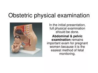
Obstetric physical examination
Obstetric physical examination. In the initial presentation, full physical examination should be done. Abdominal & pelvic examination remains important exam for pregnant women because it is the easiest method of fetal monitoring.
4.52k views • 17 slides
Introduction Physical Examination
Introduction Physical Examination. Health Assessment. Why do I need to know Physical Assessment?. “What is your reason for coming here today?”. Assessment. Collection of data about an individual’s health state. Subjective Data Client history Objective Data Inspection Percussion
1.28k views • 20 slides

Physical Examination
Physical Examination. General Considerations. General Comments. The exam should be performed in a logical manner with smooth transition from one area to the next Do not use ‘jargon’ when speaking to the patient Explain the parts of the exam to the patient
954 views • 4 slides
Physical examination
Physical examination. Body weight is often low ; asthenic Blood pressure is usually normal or low Orthostatic hypotension Thoracic skeletal abnormalities suggesting MVP: Scoliosis, pectus excavatum , straightened thoracic spine, and narrowed AP dm of the chest.
428 views • 4 slides

PHYSICAL EXAMINATION
PHYSICAL EXAMINATION. GENERAL SURVEY. Baby is awake, comfortable, no gross malformations observed and not in cardiorespiratory distress. ANTHROPOMETRIC MEASUREMENTS AND VITAL SIGNS. Head Circumference: 33 cm Chest Circumference: 29 cm Abdominal Circumference: 30 cm Length: 47 cm
622 views • 10 slides
GIT Physical Examination
GIT Physical Examination . Hadeel Khadawardi , teaching assistant at Internal Medicine Department, Faculty of Medicine, Umm Al- Qura University. Introduction . Cachectic Jaundiced . General Approach . Vital Signs . Position . Flat On one pillow . GIT Exam . Abdominal Exam .
9.32k views • 43 slides
CVS Physical Examination
CVS Physical Examination . Hadeel Khadawardi , teaching assistant at Internal Medicine Department, Faculty of Medicine, Umm Al- Qura University. General Approach . Vital Signs . Position . 45. CVS Exam . Pericardium Exam . Peripheral Exam . Inspection Palpation Auscultation . Hand
1.15k views • 24 slides
RS Physical Examination
RS Physical Examination . Hadeel Khadawardi , teaching assistant at Internal Medicine Department, Faculty of Medicine, Umm Al- Qura University. Introduction . Coughing/ Sputum Strider/ Wheezing . General Approach . Vital Signs . Position . 45 sitting over the edge of bed or
1.66k views • 46 slides
การตรวจร่างกาย Physical Examination
การตรวจร่างกาย Physical Examination. นพ . สาธิต กาสุริย์. Vital Signs. Equipment Needed General Considerations Temperature Respiration Pulse Interpretation Blood Pressure Interpretation Notes. Equipment Needed. A Stethoscope A Blood Pressure Cuff A Watch Displaying Seconds
2.6k views • 231 slides

Physical Examination: Thorax
Physical Examination: Thorax. Thorax. Heart Lungs Inspect, palpate, percuss, auscultate. Anterior Chest Landmarks. Anterior: Midsternal, midclavicular, anterior axillary
1.67k views • 47 slides
PHYSICAL EXAMINATION
PHYSICAL EXAMINATION. Afnan .Y. Toonsi. INTEGUMENTARY SYSTEM. Skin: Inspect skin color. Inspect uniformity of skin color. Assess for edema (location, color, temperature, shape, and degree.
1.03k views • 26 slides
 Preparticipation Physical Examination" width="320px" />
Preparticipation Physical Examination. Deb Jacobson, M.D. Goals and objectives. Eval general health status Determine fitness for competition Foster injury prevention Discover diseases/injuries Counsel on health-related issues. Detect conditions that may be life-threatening or disabling
775 views • 22 slides

Physical examination
Physical examination. Department of Gastroenterology Ren Ji Hospital Prof. Zhi Hua Ran. Physical examination. It is the process of examining the patient’s body to determine the presence or absence of physical problems
1.42k views • 60 slides
Introduction to the Physical Examination
Introduction to the Physical Examination. Today’s Agenda. Overview of course Exam techniques and use of equipment Vital signs. Introduction to the Medical Profession. Not an introduction, but a beginning A new type of learning experience The study of the patient
1.96k views • 124 slides

PHYSICAL EXAMINATION
PHYSICAL EXAMINATION. Examination of the ear and related head and neck structures should be performed in a systematic and consistent manner so that no part of the exam is neglected. EXTERNAL AUDITORY CANAL (EAC). composed of cartilage covered by skin
1.43k views • 93 slides

Physical Examination
Physical Examination. On admission. Upon PE. Anthropometric Measurements : Height: 157cm Weight: 74kg BMI: 30. General Survey Conscious, coherent, stretcher-borne, in cardiorespiratory distress Vital Signs BP: 200/100, supine LUE; 190/100, RUE, SBP 190, LLE, SBP 190 RLE;
1.22k views • 19 slides
General Physical Examination
General Physical Examination. Dr Preamala.G Medical Department HTJ,Seremban. DOCTORS SHOULD BE OBSERVANT,LIKE A DETECTIVE; “CONAN DOYLE” Look at the patients general appearance…at the face ,hands and body
15.07k views • 54 slides

Physical examination
Physical examination. Department of Gastroenterology Ren Ji Hospital Prof. Zhi Hua Ran. Physical examination. It is the process of examining the patient’s body to determine the presence or absence of physical problems
1.09k views • 60 slides
Physical Examination (continued)
Physical Examination (continued). Sean Ragain MD. Quick Review. So far, you’ve covered a lot of ground. Let’s look at what you’ve already seen and heard. Inspection, palpation, percussion, auscultation (not necessarily in this order). Exam is modified per patient
613 views • 41 slides

PHYSICAL EXAMINATION
PHYSICAL EXAMINATION. Midterm Systems requirements assessment -critical elements -used on care plan -includes systems -used in hosp. CRITICAL ELEMENTS. Wash hands and gather equipment Prepare assessment area Introduce yourself and state purpose
1.06k views • 20 slides
